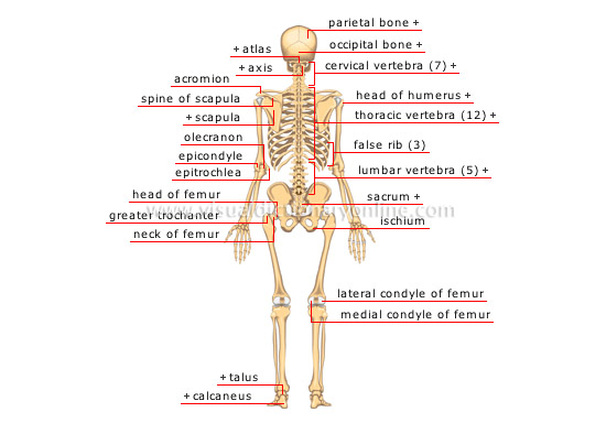
posterior view
parietal bone 
Flat cranial bone articulating with the frontal, occipital, temporal and sphenoid bones; the two parietal bones form the largest portion of the dome of the skull.
axis 
Second cervical vertebra supporting the atlas; it allows the head to rotate.
cervical vertebra (7) 
Bony part of the neck forming the upper terminal part of the spinal column.
head of humerus 
Upper terminal part of the humerus articulating very freely with the scapula.
thoracic vertebra (12) 
Bony part supporting the ribs located between the cervical and lumbar vertebrae.
false rib (3) 
Slender curved bone articulated with the dorsal vertebrae at one end and attached to the upper rib at the other end.
lumbar vertebra (5) 
Bony part larger than the other vertebrae located between the dorsal vertebrae and the sacrum; it supports a major portion of the body’s weight.
sacrum 
Bone made up of five fused vertebrae located between the lumbar and coccyx vertebrae.
lateral condyle of femur 
Round protuberance of the lower terminal part of the femur enabling articulation with the tibia.
occipital bone 
Flat skull bone articulating with the parietal bone and atlas (first cervical vertebra), among others; it makes up the largest portion of the base of the skull.
atlas 
First cervical vertebra supporting the head and supported by the axis.
acromion 
Extension of the spine of the scapula forming the point of the shoulder and articulating with the clavicle.
spine of scapula 
Pointy protuberance of the posterior scapula that extends through the acromion.
scapula 
Large thin flat bone articulating with the clavicle and the humerus to form the shoulder; numerous shoulder and back muscles are attached to it.
epicondyle 
Outer protuberance of the lower terminal part of the humerus; various extensor muscles of the hand and fingers are attached to it.
olecranon 
Upper terminal part of the ulna articulating with the humerus; it forms the protuberance of the elbow.
epitrochlea 
Inner protuberance of the lower terminal part of the humerus; various flexor muscles of the hand and fingers are attached to it.
greater trochanter 
Large protuberance of the upper terminal part of the femur; various thigh and buttock muscles are attached to it.
neck of femur 
Narrow portion of the femur connecting the head of the femur to the trochanters.
head of femur 
Upper terminal part of the femur articulating with the iliac bone to form the hip joint.
ischium 
Constituent portion of the iliac bone supporting the body’s weight when seated.
talus 
Short bone of the tarsus that, with the calcaneus, ensures rotation of the ankle and, with the tibia and fibula, flexion and extension of the foot.
calcaneus 
Large bone of the tarsus forming the protuberance of the heel; in the upright position, it is the posterior contact point of the plantar arch.


