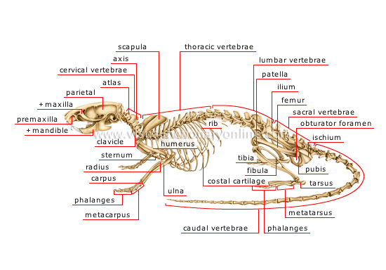
skeleton of a rat
rib 
Thin curved bone articulating with the vertebral column and the sternum.
scapula 
Large thin flat shoulder bone articulating with the humerus.
ilium 
Large flat back bone articulating with the sacral vertebrae.
patella 
Small flat slightly bulging triangular bone located on the inner limb and articulating especially with the femur.
obturator foramen 
Opening located in the lower part of the bone formed by the ilium, the ischium and the pubis; it is partly sealed by a membrane and muscles.
femur 
Long bone of the hind limb articulating especially with the patella.
pubis 
Ventral bone posterior to the ilium.
ischium 
Bone behind the ilium; the ilium, ischium and pubis fuse together to form a single bone to which the leg is attached.
phalanges 
Bones articulating to form the skeleton of the digits.
tarsus 
Part of the hind limb formed of several small bones at the juncture of the tibia and the metatarsus.
tibia 
Long bone partly fused to the fibula and forming the inner limb between the femur and the tarsus.
fibula 
Long bone partly fused to the tibia and forming the outer limb between the femur and the tarsus.
costal cartilage 
Strong elastic tissue extending the front portion of the ribs to connect them to the sternum.
sacral vertebrae 
Partly fused bony parts between the lumbar and caudal vertebrae.
thoracic vertebrae 
Bony parts supporting the ribs between the cervical and lumbar vertebrae.
caudal vertebrae 
Bony parts comprising the skeleton of the tail located at the terminal end of the vertebral column.
ulna 
Long bone partly fused with the radius and forming the inner limb between the humerus and the carpus.
radius 
Long bone partly fused with the ulna and forming the outer limb between the humerus and the carpus.
carpus 
Portion of the pectoral fin formed of short bones between the radius, the ulna and the metacarpus.
sternum 
Elongated flat bone to which the ribs in particular are attached and bearing a crest on its ventral surface.
phalanges 
Bones articulating to form the skeleton of the digits.
clavicle 
Long bone located in the front ventral portion of the body articulating with the sternum.
humerus 
Bone of the forelimb articulating with the scapula, as well as with the radius and the ulna; it provides a large base for the muscles.
atlas 
First cervical vertebra supporting the head and supported by the axis.
mandible 
Toothed bone forming the lower jaw.
axis 
Second cervical vertebra supporting the atlas; it allows the head to rotate.
lumbar vertebrae 
Bony parts of the back located between the thoracic and sacral vertebrae.
cervical vertebrae 
Bony parts of the neck comprising the upper terminal end of the vertebral column.
premaxilla 
Bone forming the anterior portion of the upper jaw.
parietal 
Flat bone of the upper portion of the skull.
maxilla 
Toothed bone forming, with the premaxilla, the upper jaw.


